A new urine diagnostic to detect the parasitic worms that cause river blindness (Onchocerciasis)
August 30th, 2018Ryan Shirey, Daniel Globisch, Lisa M Eubanks, Mark S Hixon, Kim D Janda. Non-Invasive Urine Biomarker Lateral Flow Immunoassay for Monitoring Active Onchocerciasis. ACS Infectious Diseases, 2018; DOI: 10.1021/acsinfecdis.8b00163
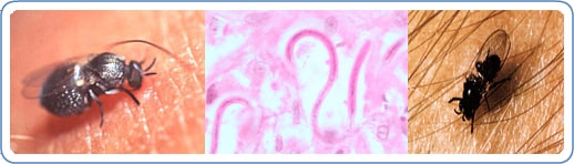
During a blood meal, an infected blackfly (genus Simulium) introduces third-stage filarial larvae onto the skin of the human host, where they penetrate into the bite wound  . In subcutaneous tissues the larvae
  develop into adult filariae, which commonly reside in nodules in subcutaneous connective tissues
  . Adults can live in the nodules for approximately 15 years. Some nodules may contain numerous male and female worms. Females measure 33 to 50 cm in length and 270 to 400 μm in diameter, while males measure 19 to 42 mm by 130 to 210 μm. In the subcutaneous nodules, the female worms are capable of producing microfilariae for approximately 9 years. The microfilariae, measuring 220 to 360 µm by 5 to 9 µm and unsheathed, have a life span that may reach 2 years. They are occasionally found in peripheral blood, urine, and sputum but are typically found in the skin and in the lymphatics of connective tissues
  . A blackfly ingests the microfilariae during a blood meal
  . After ingestion, the microfilariae migrate from the blackfly’s midgut through the hemocoel to the thoracic muscles
  . There the microfilariae develop into first-stage larvae
  and subsequently into third-stage infective larvae
  . The third-stage infective larvae migrate to the blackfly’s proboscis
  and can infect another human when the fly takes a blood meal .