CDC on NiV (Nipah virus)
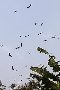
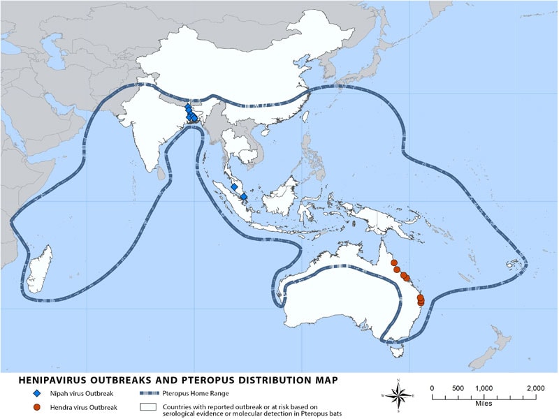
May 25th, 2018Nipah virus (NiV) is a member of the family Paramyxoviridae, genus Henipavirus. NiV was initially isolated and identified in 1999 during an outbreak of encephalitis and respiratory illness among pig farmers and people with close contact with pigs in Malaysia and Singapore. Its name originated from Sungai Nipah, a village in the Malaysian Peninsula where pig farmers became ill with encephalitis. Given the relatedness of NiV to Hendra virus, bat species were quickly singled out for investigation and flying foxes of the genus Pteropus were subsequently identified as the reservoir for NiV (Distribution Map).
In the 1999 outbreak, Nipah virus caused a relatively mild disease in pigs, but nearly 300 human cases with over 100 deaths were reported. In order to stop the outbreak, more than a million pigs were euthanized, causing tremendous trade loss for Malaysia. Since this outbreak, no subsequent cases (in neither swine nor human) have been reported in either Malaysia or Singapore.
In 2001, NiV was again identified as the causative agent in an outbreak of human disease occurring in Bangladesh. Genetic sequencing confirmed this virus as Nipah virus, but a strain different from the one identified in 1999. In the same year, another outbreak was identified retrospectively in Siliguri, India with reports of person-to-person transmission in hospital settings (nosocomial transmission). Unlike the Malaysian NiV outbreak, outbreaks occur almost annually in Bangladesh and have been reported several times in India.
Transmission

Transmission of Nipah virus to humans may occur after direct contact with infected bats, infected pigs, or from other NiV infected people.
In Malaysia and Singapore, humans were apparently infected with Nipah virus only through close contact with infected pigs. The NiV strain identified in this outbreak appeared to have been transmitted initially from bats to pigs, with subsequent spread within pig populations. Incidental human infections resulted after exposure to infected pigs. No occurrence of person-to-person transmission was reported in this outbreak.
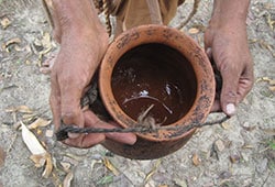
Conversely, person-to-person transmission of Nipah virus in Bangladesh and India is regularly reported. This is most commonly seen in the family and caregivers of Nipah virus-infected patients. Transmission also occurs from direct exposure to infected bats. A common example is consumption of raw date palm sap contaminated with infectious bat excretions.
Signs and Symptoms
Infection with Nipah virus is associated with encephalitis (inflammation of the brain). After exposure and an incubation period of 5 to 14 days,illness presents with 3-14 days of fever and headache, followed by drowsiness, disorientation and mental confusion. These signs and symptoms can progress to coma within 24-48 hours. Some patients have a respiratory illness during the early part of their infections, and half of the patients showing severe neurological signs showed also pulmonary signs.
During the Nipah virus disease outbreak in 1998-99, 265 patients were infected with the virus. About 40% of those patients who entered hospitals with serious nervous disease died from the illness.
Long-term sequelae following Nipah virus infection have been noted, including persistent convulsions and personality changes.
Latent infections with subsequent reactivation of Nipah virus and death have also been reported months and even years after exposure.
Risk of Exposure

In the Malaysia and Singapore outbreak, Nipah virus infection was associated with close contact with Nipah virus-infected pigs.
In Bangladesh and India, where Nipah virus infection is more frequent, exposure has been linked to consumption of raw date palm sap and contact with bats. Importantly, human-to-human transmission has been documented and exposure to other Nipah virus infected individuals is also a risk factor.
Diagnosis

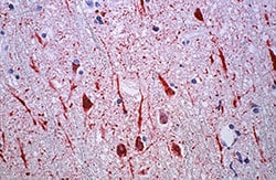
Laboratory diagnosis of a patient with a clinical history of NiV can be made during the acute and convalescent phases of the disease by using a combination of tests. Virus isolation attempts and real time polymerase chain reaction (RT-PCR) from throat and nasal swabs, cerebrospinal fluid, urine, and blood should be performed in the early stages of disease. Antibody detection by ELISA (IgG and IgM) can be used later on. In fatal cases, immunohistochemistry on tissues collected during autopsy may be the only way to confirm a diagnosis.
Treatment
Treatment is limited to supportive care. Because Nipah virus encephalitis can be transmitted person-to-person, standard infection control practices and proper barrier nursing techniques are important in preventing hospital-acquired infections (nosocomial transmission).
The drug ribavirin has been shown to be effective against the viruses in vitro, but human investigations to date have been inconclusive and the clinical usefulness of ribavirin remains uncertain.
Passive immunization using a human monoclonal antibody targeting the Nipah G glycoprotein has been evaluated in the post-exposure therapy in the ferret model and found to be of benefit.
Prevention

Nipah virus infection can be prevented by avoiding exposure to sick pigs and bats in endemic areas and not drinking raw date palm sap.
Additional efforts focused on surveillance and awareness will help prevent future outbreaks. Research is needed to better understand the ecology of bats and Nipah virus, investigating questions such as the seasonality of disease within reproductive cycles of bats. Surveillance tools should include reliable laboratory assays for early detection of disease in communities and livestock, and raising awareness of transmission and symptoms is important in reinforcing standard infection control practices to avoid human-to-human infections in hospital settings (nosocomial infection).
A subunit vaccine, using the Hendra G protein, produces cross-protective antibodies against HENV and NIPV has been recently used in Australia to protect horses against Hendra virus. This vaccine offers great potential for henipavirus protection in humans as well.

References
- MMWR, Outbreak of Hendra-like virus—Malaysia and Singapore, 1998-1999. Apr 9, 1999;48(3):265-9.
- MMWR, Update: Outbreak of Nipah virus– Malaysia and Singapore, 1999. Apr 30, 1999;48(16):335-7.
- Broder CC. Henipavirus outbreaks to antivirals: the current status of potential therapeutics. Current Opinion Virology 2012;2(2):176-87.
- Chadha MS, Comer JA, Lowe L, et al. Nipah virus-associated encephalitis outbreak, Siliguri, India. Emerging Infectious Disease 2006;12(2):235-40.
- Chua KB, Goh KJ, Wong KT, et al. Fatal encephalitis due to Nipah virus among pig-farmers in Malaysia. Lancet 1999;354(9186):1257-9.
- Chua KB, Bellini WJ, Rota PA, et al. Nipah virus: A recently emergent deadly paramyxovirus. Science 2000;288(5470):1432-5.
- Chua KB, Lam SK, Goh KJ, et al. The presence of Nipah virus in respiratory secretions and urine of patients during an outbreak of Nipah virus encephalitis in Malaysia. Journal of Infection 2001;42(1):40-3.
- Daniels P, Ksiazek T, Eaton BT. Laboratory diagnosis of Nipah and Hendra virus infections. Microbes and Infection 2001;3(4):289-95.
- Field HE, Mackenzie JS, Daszak P. Henipaviruses: emerging paramyxoviruses associated with fruit bats. Current Topics Microbiology and Immunology 2007;315:133-59.
- Gurley ES, Montgomery JM, Hossain MJ, et al. Person-to-person transmission of Nipah virus in a Bangladeshi community. Emerging Infectious Disease 2007;13(7):1031-7.
- Hossain MJ, Gurley ES, Montgomery JM, et al. Clinical presentation of Nipah virus infection in Bangladesh. Clinical Infectious Diseases 2008;46(7):977-84.
- Lee KE, Umapathi T, Tan CB, et al. The neurological manifestations of Nipah virus encephalitis, a novel paramyxovirus. Annals of Neurology 1999;46428-32.
- Lim CCT, Lee KE, Lee WL, et al. Nipah virus encephalitis: Serial MR study of an emerging disease. Radiology 2002;222(1):219-26.
- Luby SP, Gurley ES, Hossain MJ. Transmission of human infection with Nipah virus. Clinical Infectious Disease 2009;49(11):1743-8.
- Mounts AW, Kaur H, Parashar UD, et al. A cohort study of health care workers to assess nosocomial transmissibility of Nipah virus, Malaysia, 1999. Journal of Infectious Disease 2001;183(5):810-3.
- Murray K, Selleck P, Hooper P, et al. A morbillivirus that caused fatal disease in horses and humans. Science 1995;268:94-7.
- Paton NI, Leo YS, Zaki SR, et al. Outbreak of Nipah-virus infection among abattoir workers in Singapore. Lancet 1999;354(9186):1253-6.
- Rahman MA, Hossain MJ, Sultana S, et al. Date Palm Sap Linked to Nipah Virus Outbreak in Bangladesh, 2008. Vector-Borne and Zoonotic Disease 2012;12(1):65-73.
- Reynes J-M, Counor D, Ong S, et al. Nipah virus in Lyle’s Flying Foxes, Cambodia. Emerging Infectious Disease 2005;11(7):1042-7.
- Rollin PE, Rota P, Zaki S, Ksiazek TG. Hendra and Nipah viruses. in: Versalovic J, Carroll KC, Funke G, Jorgensen JH, Landry ML, Warnock DW, editors. Manual of Clinical Microbiology. 10th ed. Washington, DC: ASM Press; 2011; p. 1479-87.
- Sim BF, Jusoh MR, Chang CC, Khalid R. Nipah Encephalitis: A report of 18 patients from Kuala Lumpur Hospital. Neurology Journal Southeast Asia 2002;7:13-8.
- Tan CT, Goh KJ, Wong KT, et al. Relapsed and Late-Onset Nipah Encephalitis. Ann. Neurology 2002;51(6):703-8.
- Wacharapluesadee S, Boongird K, Wanghongsa S, et al. A Longitudinal Study of the Prevalence of Nipah Virus in Pteropus lylei Bats in Thailand: Evidence for Seasonal Preference in Disease Transmission. Vector-Borne and Zoonotic Disease 2010;10(2):183-90.
- Williamson M, Torres-Velez FJ. Henipavirus: a review of laboratory animal pathology. Veterinary Pathology 2010;47(5):871-80.
- Wong KT, Shieh WJ, Kumar S, et al. Nipah virus infection. Pathology and pathogenesis of an emerging paramyxoviral zoonosis. American Journal of Pathology 2002;161(6):2153-67.

