Archive for the ‘Thelaziasis’ Category
Thelaziasis image gallery
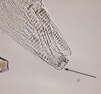
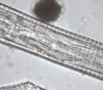
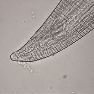
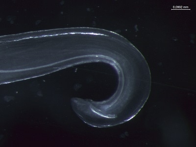
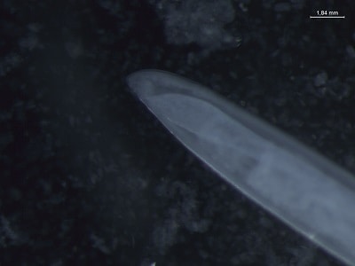
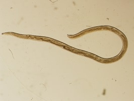
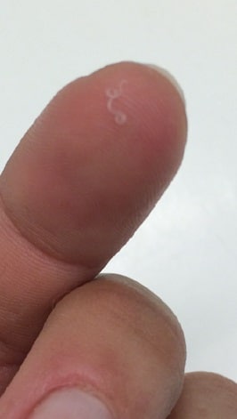
Tuesday, November 12th, 2019Thelazia spp. adults.Adults of Thelazia spp. reside in the conjunctival sac of their definitive hosts, which are usually dogs or other canids, cattle, and horses; humans are usually only incidental hosts. Adults measure up to 2.1 cm in length. Thelazia species may be differentiated by the appearance of the cuticular striations, the depth and width of the buccal cavity, the placement of the vulval opening relative to the esophago-intestinal junction and the morphology of the tail and anal opening.
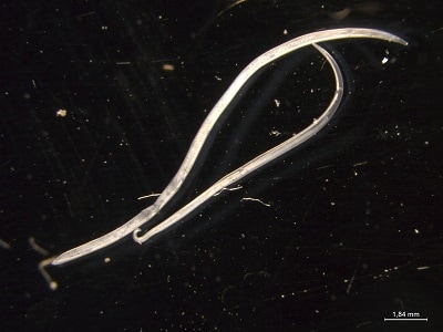
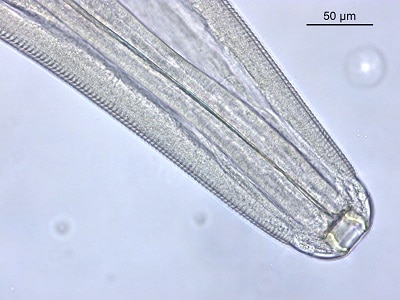
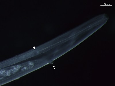
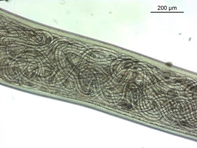


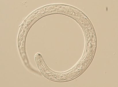
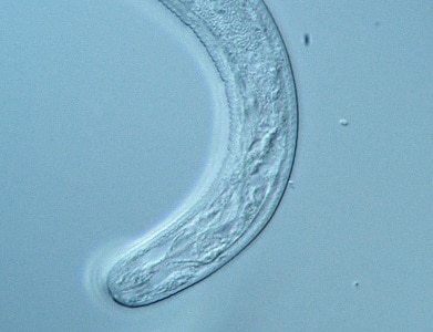
Thelaziasis
Wednesday, November 6th, 2019Causal Agents
Spirurid nematodes in the genus Thelazia are primarily veterinary parasites, but may occasionally infect humans. The majority of zoonotic infections involve T. callipaeda (the Oriental eye worm). T. californiensis (the California eye worm) and T. gulosa (the cattle eyeworm) are less common causative agents.
Life Cycle
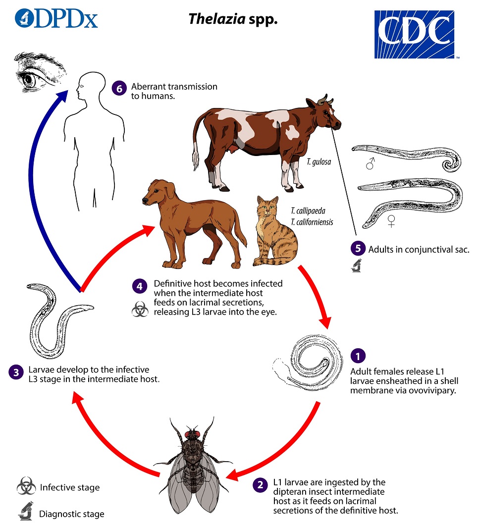
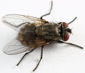
Adults reside in the conjunctival sac of the definitive host where the ovoviviparous females release first-stage (L1) larvae ensheathed in a shell membrane  . L1 larvae are ingested by the face fly intermediate host during feeding on tears and lacrimal secretions
  . In the digestive tract of the intermediate host, L1 larvae become exsheathed and invade various host tissues, including the hemocoel, fat body, testis, and egg follicles where they develop in capsules. The encapsulated larvae molt twice to become infective L3 larvae
  . The fully developed L3 larvae break out of the capsules and migrate to the fly’s mouthparts, where they remain until the fly feeds on the tears of the definitive host. The larvae invade the conjunctival sac of the definitive host upon the fly intermediate host’s feeding
  and become adults after about a month and two additional molts
  . Humans may also serve as aberrant definitive hosts following exposure to an infected fly intermediate host in the same manner
  .
.
Hosts and Vectors
Both wild and domestic canids are considered the primary definitive hosts for Thelazia callipaeda. Natural infections have also been detected in felids, mustelids, and lagomorphs. T. californiensis infections have been reported in numerous mammals, mostly from wild and domestic canids, but also others (e.g. felids, bears, cervids, sheep, jackrabbits (Lepus californicus)). Some of these could possibly represent misidentifications of species. T. gulosa is a parasite of cattle, and occasionally other large ruminants.
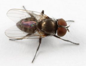
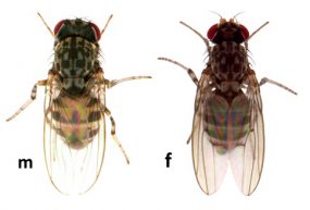
Intermediate hosts or vectors for Thelazia spp. are drosophilid flies that feed on lacrimal secretions (lacrimophagous), and associations are parasite-specific. For example, Phortica (=Amiota) variegata and P. (=A.) okadai are the primary intermediate hosts for T. callipaeda. Fannia spp., such as Fannia benjamini (canyon fly) and F. canicularis (lesser house fly), are vectors for T. californiensis. Musca autumnalis (face fly) appears to be the most important vector for T. gulosa.
Geographic Distribution
Thelazia callipaeda is widespread across Asia and has now become established in continental Europe. T. californiensis has been reported from the western United States, although characterization of its range is incomplete. T. gulosa has a wide distribution across Asia, Europe, North America, and Australia.
Clinical Presentation
Adults in the eye cause varying degrees of inflammation and lacrimmation accompanied by a foreign body sensation. . In heavier infections, photophobia, epiphora, edema, corneal ulceration, and conjunctivitis may occur.


